Japan (Kyushu University) 3D flora and fauna at your fingertips
Reporting in Research Ideas and Outcomes, a Kyushu University researcher has developed a new technique for scanning various plants and animals and reconstructing them into highly detailed 3D models. To date, over 1,400 models have been made available online for public use.
Open any textbook or nature magazine and you will find stunning high-resolution pictures of the diverse flora and fauna that encompass our world. From the botanical illustrations in Dioscorides’ De materia medica (50–70 CE) to Robert Hooke’s sketches of the microscopic world in Micrographia (1665), scientists and artists alike have worked meticulously to draw the true majesty of nature.
The advent of photography has given us even more detailed images of animals and plants both big and small, in some cases providing new information on an organism’s morphology. As technology developed, digital libraries began to grow, giving us near unfettered access to valuable data, with methods like computer tomography, or CT, and MRI scanning becoming powerful tools for studying the internal structure of such creatures.
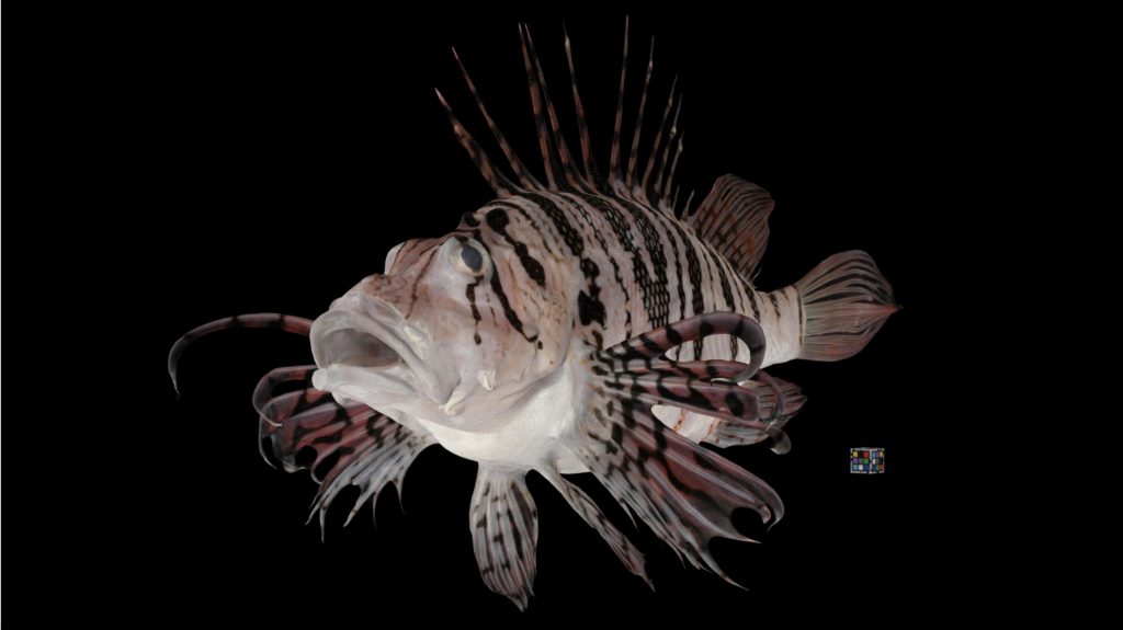
Luna Lionfish, Pterois lunulate. 3D render of a Luna Lionfish taken with bio-photogrammetry
Luna Lionfish, Pterois lunulate. 3D render of a Luna Lionfish taken with bio-photogrammetry (Kano Lab/Kyushu University)
“While powerful, MRI scanning and CT methods are prohibitively expensive. You also can’t collect vital information such as the organism’s color,” explains Yuichi Kano, associate professor of Kyushu University’s Graduate Education and Research Training Program in Decision Science for a Sustainable Society. “So, we developed ‘bio-photogrammetry’ as a way to incorporate photogrammetry that could scan and render a high-quality 3D image of an organism.”
Photogrammetry is a method by which you can obtain information and measurements about objects by analyzing photos or other imagery. Today it is commonly used to scan everything from landscapes to sculptures to make digital 3D models, similar to what you find on Google Earth.
Kano took that same methodology to make thousands of models of various organisms.
“We suspended the sample on a fishing line and took photos from multiple angles. We would end up taking hundreds of photos of the sample, and input up to 500 of the best ones into the photogrammetry program,” explains Kano. “It is similar to how the ‘bullet time’ sequences were filmed in the first Matrix movie, except instead of Keanu Reeves on a line surrounded by cameras, we use an octopus.”
While Kano has been working on various organisms including insects, plants, and even fungi, he is currently focusing on aquatic animals such as fish and amphibians. To date, there are over 1,400 specimens available all free to use under the CC BY 4.0 license.
There are a few limitations in the current methodology, such as difficulty in capturing transparent creatures or making models of exceedingly small (<5 mm) or large (>1 m) organisms, but a few improvements in software and protocols could help solve such issues.
“I hope to see this work continue to grow and be utilized in various fields like taxonomy, morphology, and ecology. It’s free to the public, so you can use it in education or even plug it into a VR machine and explore these organisms up-close. I’d like to see what some people can come up with,” concludes Kano.